
Dr. Victoria P. Werth explains when and why invasive muscle biopsies might not be necessary in classic dermatomyositis with diagnostic skin findings and suggests less invasive and less costly alternatives.
“When patients have dermatomyositis and muscle complaints—even if they have a skin rash—they often end up with a muscle biopsy. You really don’t need to do a muscle biopsy in patients who have interface dermatitis and skin findings that look like dermatomyositis,” said Victoria P. Werth, MD, senior author of the study “Diagnosing muscle disease in a cohort of classic dermatomyositis patients seen at a rheumatologic dermatology outpatient clinic,” published March 1, 2022, in the Journal of the American Academy of Dermatology (JAAD).1
Dr. Werth and coauthors reviewed the cases of 275 dermatomyositis patients to determine whether skin biopsy, electromyography (EMG), or magnetic resonance imaging (MRI) of the involved muscle could be done instead of muscle biopsy. They found that 65% of the muscle biopsy cases with findings consistent with dermatomyositis were in agreement with diagnostic features on EMG or MRI. Skin and muscle biopsy results supported dermatomyositis in 67% of patients who underwent both procedures.
“In the presence of [dermatomyositis]-specific skin findings, less invasive procedures may be sufficient to diagnose [dermatomyositis] and guide its management,” the authors conclude.
“Muscle biopsy is a fairly morbid procedure. Patients complain about it for a long time after. And this study shows that it’s not necessary in many cases,” said Dr. Werth.
While rheumatologists are more likely than dermatologists to use muscle biopsies to diagnose muscle symptoms associated with dermatomyositis, dermatologists play a role in making the diagnosis based on skin findings, avoiding the need for a muscle biopsy, said Dr. Werth.
“I don’t want to make it sound like there is never a reason for a muscle biopsy. There are patients who have only muscle disease and there are other things that could be causing their muscle symptoms. Those kinds of patients go to rheumatologists and neurologists—we don’t see them in dermatology. Those patients need a muscle biopsy.”
Diagnostic Alternatives
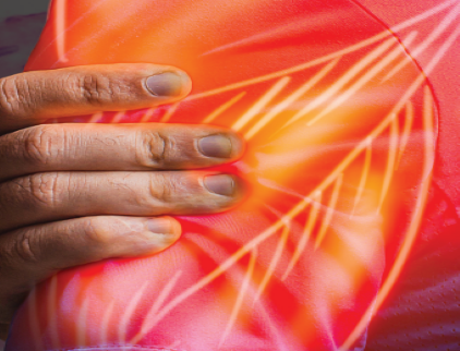
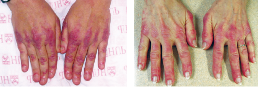
Rather, Dr. Werth is referring to patients with classic dermatomyositis skin findings, such as Gottron papules, Gottron sign, and heliotrope rash.
Options for diagnosing those patients include a skin biopsy and testing for autoantibodies and muscle enzymes with the creatine phosphokinase (CPK) aldolase.
“We can also sometimes see elevated transaminases, which can be coming from the muscle. Often, people are getting worked up for liver problems when they actually have a muscle disease, and the enzymes are coming from the muscle.”
While cutaneous findings, muscle symptoms consistent with dermatomyositis and a skin biopsy consistent with dermatomyositis, should be sufficient to establish the diagnosis, that doesn’t rule out the need for an EMG or MRI, said Dr. Werth.
“We often [treat with] systemic medication, whether it be steroids or immunosuppressives, and so on, so you want to document that there is indeed muscle involvement.”
“An EMG or MRI can be quite helpful in making a diagnosis. Not that they’re perfect, but they can be very helpful,” she said.
Muscle biopsies, according to the study, also are not perfect. Eight of 21 patients studied with a clinical diagnosis of dermatomyositis but negative muscle biopsies had EMG or MRI with results that were consistent with the diagnosis, suggesting that relying only on a muscle biopsy is not enough.
Consider This Diagnostic Algorithm
Dr. Werth and coauthors suggested a simple algorithm for diagnosing dermatomyositis in patients with skin findings.
First, determine if the patient has a rash that is characteristic of dermatomyositis, including erythema over the joints, eyelids, face and often the nasolabial fold, upper back, and upper arms. Sometimes patients will have nailfold capillary changes.
With skin findings that are consistent with dermatomyositis, Dr. Werth said, “if there is an area that you can biopsy, and often there is, then doing the skin biopsy would be helpful to rule out other mimickers.”
Skin findings and a skin biopsy that is consistent with dermatomyositis can lead to the diagnosis.
“I would say about 80% of the time a skin biopsy will show interface dermatitis. Some skin biopsies—especially those with more damage than an activity—do not show interface dermatitis. But that doesn’t mean that the patient doesn’t have dermatomyositis.”
Another potential hurdle in making the diagnosis is that dermatopathologists often report interface dermatitis as being consistent with lupus, according to Dr. Werth.
“What ends up happening is the biopsy result will flow to primary care or to rheumatology and the biopsy reports the patient has lupus. They don’t necessarily know how to look at the skin, so it really requires a dermatologist to understand that the interface dermatitis seen on the biopsy could be seen in either lupus or dermatomyositis. And if the clinical findings look like dermatomyositis, it is dermatomyositis.”
If the patient also has proximal weakness or pain, then Dr. Werth recommends checking CPK aldolase.
“Even if [the CPK aldolase is] normal, you’re still going to want a patient who has muscle findings and symptoms to get an MRI or EMG to document whether muscle disease can be documented.”
Whether to choose an EMG or MRI depends on patient preferences and access, according to Dr. Werth.
“EMG requires some expertise and may not be readily available. You need a neurologist who is experienced to interpret the results. Some patients don’t like to have needles stuck in them. But the advantage can be that you can sample more muscle groups.”
“If you can target a muscle group, then an MRI would be helpful. You can’t MRI the whole body, so you have to be able to say which muscle group you want to image based on pain and/or weakness. In general, I think people tolerate MRIs better than EMGs.”
In the study, the cost difference between MRI and EMG was not significant, but the cost of muscle biopsies was nearly 10 times higher than that of the other two tests.
“Normal findings [on an EMG or MRI] will guide your therapy. You’ll be able to say that patient has classic dermatomyositis, and you would probably initially start more aggressive therapy, which would likely include systemic steroids and other steroid-sparing approaches.”
The myositis community, especially rheumatologists that specialize in myositis, would likely agree with this approach outlined in the study, according to Dr. Werth.
“The problem really comes down to educating people beyond the specialists who focus on myositis, meaning general rheumatologists and dermatologists, in terms of trying to make sure that it’s not a knee-jerk reaction to order a muscle biopsy just because a patient has muscle symptoms. We are trying to make people more aware that you don’t have to have a muscle biopsy in the setting where you have the skin findings and the skin biopsy to confirm the diagnosis of dermatomyositis.”
Reference:
- Ahmed S, Concha JSS, Chakka S, et al. Diagnosing muscle disease in a cohort of classic dermatomyositis patients seen at a rheumatologic dermatology outpatient clinic. J Am Acad Dermatol. 2022;86(3):544-550. doi:10.1016/j.jaad.2021.05.026
Disclosures: Dr. Werth reports no relevant disclosures.
