Researchers have developed an index of the threshold energy density, known as fluence, and the dependent wavelength for picosecond lasers to aid spot treatment of pigmented lesions.
For the study, researchers developed threshold fluences for melanosome disruption with picosecond lasers by evaluating the relationship between the irradiation fluence and mean melanosome particle size and confirming melanosome disruption by scanning electron microscope when a significant change in the mean particle size was observed. The threshold fluences were determined to be 0.95, 2.25, 2.75, and 6.50 J/cm² for 532-, 730-, 785-, and 1064-nm picosecond lasers, respectively, the study showed.
Comparing previously reported clinical studies, the researchers confirmed that clinical results showing low complication rates and high efficacy can be explained based on these wavelength-dependent indicators.
“The use of this indicator is expected to play an important part in setting irradiation conditions in clinical practice,” says Postdoctoral Fellow Yu Shimojo of Osaka Metropolitan University’s Graduate School of Medicine in Japan, in a news release. “In addition, the implementation of picosecond laser therapy based on scientific evidence, rather than relying solely on physicians’ experience, is expected to improve the safety and effectiveness of the treatment.”
The findings were published in Lasers in Surgery and Medicine.
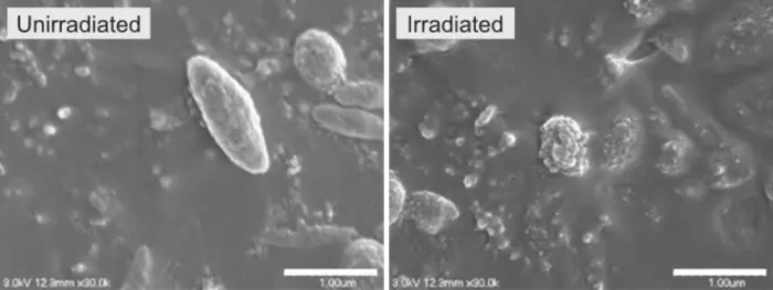
PHOTO CAPTION: Left: Unirradiated melanosomes show a smooth surface. Right: Irradiation by picosecond lasers with a pulse width of 450 ps at a wavelength of 1064 nm and a fluence of 8.50 J/cm² results in melanosomes with their surface structure disrupted.
PHOTO CREDIT: Osaka Metropolitan University
