A newly developed diagnostic system uses artificial intelligence (AI) to accurately identify the type of facial pigmented lesions and support treatment decisions as appropriate therapy for such lesions varies greatly depending on the type.
The system demonstrated superior diagnostic accuracy compared to dermatologists for identifying five types of lesions that can be difficult to diagnose, according to research published in Cureus.
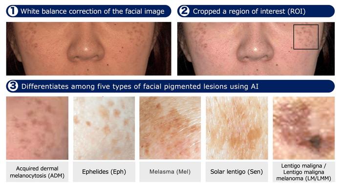
Researchers from Kindai University in Higashi-osaka, Japan, and other instotutions developed a classification system using deep learning models, InceptionResNetV2 and DenseNet121, to identify five types of facial pigmented lesions (melasma, ephelides, acquired dermal melanocytosis, solar lentigo, and lentigo maligna/lentigo maligna melanoma). Training and validation were performed using 432 clinical images, and their diagnostic accuracies were compared to the diagnoses of nine board-certified dermatologists and 11 non-experts.
Both models demonstrated diagnostic accuracies of 87% and 86%, respectively. In addition, both models overperformed the median diagnostic accuracy of 80% for experts and 63% for non-experts, the study showed. Voth models achieved 100% sensitivity, especially in identifying lentigo maligna/lentigo maligna melanoma, suggesting its potential use as a diagnostic support tool in clinical practice.
PHOTO CAPTION: A typical image preprocessing workflow. The facial image is corrected for white balance with Python’s OpenCV. A square region of interest (ROI) containing the main lesion is cropped. ROIs from five lesion types (ADM, Eph, Mel, Sen, LM/LMM) still show natural variation in skin tone and pigmentation. Only images with sufficient clarity (Laplacian variance ≥ 10) proceed for CNN analysis.
PHOTO CREDIT: Professor Atsushi Otsuka from Kindai University Faculty of Medicine, Japan
