
With Curtis T. Thompson, MD, and Eugene Semenov, MD

To avoid missing a nail-unit melanoma, experts advise knowing telltale signs and who is at risk. Effective treatment demands a thorough approach to biopsy, differential diagnosis, and therapy, they add, while avoiding overtreatment.
Since amputation of nail units or entire digits can substantially affect patients’ daily functioning, it is crucial to distinguish benign melanocytic lesions from melanoma in situ and invasive melanoma,1 according to Curtis T. Thompson, MD, Affiliate Clinical Professor of Dermatology and Pathology at Oregon Health and Sciences University in Portland, Oregon.
Acral melanomas (also called acral lentiginous melanomas) occur on the palms, soles, ears, or in the nail bed and account for 2% to 3% of all melanomas.2 Patients are typically in their 60s when diagnosed, said Eugene Semenov, MD, Instructor in the Department of Dermatology at Massachusetts General Hospital and Harvard Medical School, Boston.
Although classic cutaneous melanomas typically occur in sun-exposed Caucasian skin, said Dr. Semenov, acral melanoma is not UV-related and can affect all races, with a disproportionate burden in skin of color.3 However, he added that because acral melanomas appear in easily overlooked areas and are commonly misdiagnosed, they are often diagnosis in advanced stage.4 Subungual melanoma is considered a clinical subtype of acral melanoma, noted Dr. Semenov.
However, Dr. Thompson noted that acral and subungual melanomas probably do not share a common etiology. “There are 2 types of melanomas in the nail,” he said.
Dr. Thompson noted that the more common benign type occurs on the epithelium without invading, similar to melanoma in situ; the second, an invasive type of nail-unit melanoma, is probably more closely related to acral melanoma. “We don’t know whether either of these comes from the same source as acral melanoma, but I would speculate that the invasive type does,” he said, adding that the invasive variety occurs in a broader age spectrum, starting around age 35.
Risk factors for acral melanoma include burns and repetitive trauma, such as that which occurs with heavy machinery or hand tools, said Dr. Semenov. “But understanding of the risk factors for this disease is very limited due to the absence of rigorous studies with robust control populations,” he said.
The differential diagnosis for pigmented nail lesions is broad and includes benign nail matrix nevi, nail lentigo, longitudinal melanonychia, subungual hematoma, onychomycosis, onychopapilloma, and even squamous cell carcinoma, said Dr. Semenov.4
Nail-unit pigment also may stem from medications, Dr. Thompson said. The lesion that was historically called a benign melanotic macule or benign lentigo of the nail unit is now called benign activation of junctional melanocytes, he added. Its clinical calling card is newly developed black or brown longitudinal melanonychia. Biopsies of such areas show a slightly elevated number of melanocytes, said Dr. Thompson, although these lesions remain benign.
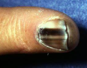
One of the key clinical features, Dr. Semenov noted, is whether a pigmented lesion involves the proximal nail fold (Hutchinson’s sign). “If it involves the proximal nail fold, our suspicion for nail melanoma increases,” he said. Other important clinical features include the width of the pigmented band, pigment irregularity, nail dystrophy, and evolution. Specifically, he said, a hallmark of concerning pigmented nail lesions is that as the nail plate grows out, the lesion does not.
The optimum biopsy technique for suspected fingernail- or toenail-unit melanomas may depend on whom one asks. “The best way is for the nail plate to be reflected back,” said Dr. Thompson, “and then for the very delicate nail matrix and nail bed epithelium to be shave biopsied and submitted very carefully to preserve the tissue integrity.”5
Conversely, Dr. Semenov recommended a punch biopsy of the nail matrix performed by a dermatologist or hand surgeon, although horizontal elliptical excisions are also used. “We perform the biopsy right over the lunula, between the proximal and distal nail matrix. For punch biopsies, a 2- to 4-mm biopsy over the affected lesion is performed and often left to heal by second intent. For excisional and elliptical biopsies or biopsies of larger size, closure with sutures is often necessary. In general, these are very uncomfortable procedures and typically require digital blocks for anesthesia,” he said, adding that these procedures may also result in permanent nail scarring.
Whichever biopsy technique is used, said Dr. Thompson, the combination of hard nail plate and delicate nail matrix/bed makes laboratory processing of specimens difficult and inconclusive results common. The challenging hard and soft composition of nail-unit tissue also explains why many clinicians are not comfortable performing nail-unit biopsies, he noted.
Nevertheless, said Dr. Thompson, dermatologists should not fear biopsying pigmented nail lesions. “They have the tools, and it’s worthwhile, if you don’t have easy access to a nail expert, to do the biopsy yourself, following the submission guidelines.”5
However, Dr. Thompson cautions against biopsying pigmented nail lesions in children. Worried parents often push for biopsies, he said. But because reliable molecular tools for detecting malignant transformation in pigmented nail lesions are lacking,6 analysis often proves inconclusive. Additionally, melanoma in children is exceedingly rare. Dr. Thompson therefore recommends referring children with suspected nail-unit melanomas to nail experts.
As with melanomas elsewhere on the body, treatment of nail-unit melanomas depends on stage at diagnosis, according to Dr. Semenov. “That is largely determined by the Breslow depth, the depth of invasion from the basal layer of the epidermis into the dermis,” he said.7
The extent of Breslow depth may warrant additional workup, he said. “Anything less than 1 mm, as long as it doesn’t have high-risk features like ulceration or presence of mitoses, we classify as T1a.7 The treatment there would be to cut out the melanoma. If it’s a digital or subungual melanoma, it may result in amputation of the distal extremity.” Fortunately, Dr. Thompson added, the trend among clinical dermatologists is toward clear-margin excision, avoiding amputations because they do not appear to improve survival.3,8
If the melanoma extends deeper than 1 mm, Dr. Semenov said, clinicians typically offer a sentinel lymph node biopsy.9 “If the lymph nodes are positive, then the melanoma becomes upstaged. At that point you would do an imaging test to see if melanoma is present elsewhere in the body,” he said.
If distant metastases exist, Dr. Semenov said, patients require chemotherapy, targeted therapy, or immunotherapy. Immune checkpoint inhibitors such as ipilimumab, pembrolizumab, and nivolumab are novel immunotherapies that have significantly extended survival in patients with advanced melanoma. However, said Dr. Semenov, attempting to predict which patients will respond to immunotherapy is of considerable importance as these medications are also associated with highly morbid and potentially life-threatening adverse events.
By John Jesitus
REFERENCES:
1. Thompson CT. Evaluation of a pigmented lesion in the nail unit: how not to miss melanoma. DERMfoot 2021 Virtual Seminar; April 16, 2021.
2. Bradford PT, Goldstein AM, McMaster ML, Tucker MA. Acral lentiginous melanoma: incidence and survival patterns in the United States, 1986-2005. Arch Dermatol. 2009;145(4):427-434.
3. Desai A, Ugorji R, Khachemoune A. Acral melanoma foot lesions. part 1: epidemiology, aetiology, and molecular pathology. Clin Exp Dermatol. 2017; 42: 845-848.
4. Semenov ER. Acral melanoma clinical update. DERMfoot 2021 Virtual Seminar; April 17, 2021.
5. Reinig E, Rich P, Thompson CT. How to submit a nail specimen. Dermatol Clin. 2015;33(2):303-307.
6. Chu A, André J, Rich P, Leachman S, Thompson CT. Immunohistochemical characterization of benign activation of junctional melanocytes and melanoma in situ of the nail unit. J Cutan Beta Pathol. 2019;46(7):479-483. 7. Keung EZ, Gershenwald JE. Th
