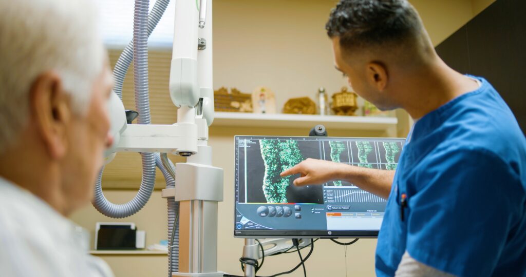
Full dermal visualization (FDV) via high-resolution dermal ultrasound (HRDUS) prior to every fraction during treatment of nonmelanoma skin cancer (NMSC) with image-guided superficial radiation therapy (Image-Guided SRT) boosts efficacy and reduces toxicity, according to a new study in Dermato.
The study involved retrospective review of the medical records of 883 patients with 1,507 cases of NMSC that were treated with Image-Guided SRT at seven dermatology clinics between 2017 and 2018.
Researchers found that 92% of NMSC tumors undergoing Image-Guided SRT exhibited measurable changes in the depth of invasion compared to that of the previous image. These measurements were collected immediately prior to treatment, which allowed the opportunity for adaptive changes in treatment parameters, such as kilovoltage (kV) x-rays, time, dose, and fractionation (TDF), dose and boost.
“These adaptive changes were necessary in nearly 40% of reviewed cases, directly benefiting patients by maximizing efficacy and minimizing toxicity,” says study author Jeffrey Stricker, DO, of Dermatology Specialists of Alabama in Huntsville, AL.
The authors emphasized that “any measured increase in NMSC depth is clinically significant during Image-Guided SRT because it lowers the percent of the prescription dose received by the tumor cells as a function of their depth (PDD). A lower PDD means significantly less radiation is delivered to the deepest tumor cells, diminishing therapeutic efficacy, potentially allowing the deepest cells to receive significantly less than required for cure. Thus, by performing HRDUS depth measurements prior to every fraction, dermatologists and RTTs know exactly when to make adaptive increases in kV, TDF and dose/boost. This ensures that every NMSC tumor is adequately and uniformly radiated to achieve 99% cure rates.”
The guiding principle of radiation safety is ALARA, which stands for “as low as reasonably achievable,” or avoiding exposure to radiation that does not have a direct benefit to the patient.
Routine HRDUS depth measurements prior to each fraction allows dermatologists and radiation therapists (RTTs) to know exactly when to make adjustments, ensuring that the safety of every NMSC patient is optimized by reducing radiation toxicity whenever possible.
The authors also noted that HRDUS imaging before each treatment session allows clinicians to monitor the gradual repopulation of a tumor and gauge the biological effectiveness of Image-Guided SRT therapy. This visualization additionally facilitates monitoring for radioresistance within a tumor, which is a possibility in the absence of the normal, expected repopulation pattern on HRDUS.
“This new study reaffirms what our partner dermatologists and Mohs surgeons have seen in their practices over the past several years, that image-informed treatments produce optimal clinical outcomes,” adds Kerwin Brandt, Chief Executive Officer of SkinCure Oncology. “We are encouraged by the growing adoption of this technology, the future of imaging in diagnostics and treatment, and the continued impact that this model of care is having on patients and their families.”


