DISCUSSION
Localization to the acral areas and a presentation that could resemble a variety of papulosquamous disorders led us to formulate a broad differential diagnosis.
Conditions such as psoriasis in skin of color, necrolytic acral erythema (NAE), nutritional deficiency, acrokeratosis paraneoplastica (Bazex syndrome), and others could be considered. Many of these manifest as a psoriasiform dermatitis. Therefore, additional work-up should be considered. Some can be associated with underlying health conditions such as malignancy and infections.
Epidemiology of NAE
NAE is a rare condition that was first described only approximately 25 years ago in a patient in Egypt.1 As of 2019, 145 cases of NAE were reported in the literature, 119 from Egypt.2 Its true prevalence is unknown, but reports have been increasing, perhaps because growing awareness is leading to increased recognition.
Most cases of NAE have been in adults who are middle aged (35 to 55 years), but affected patients have ranged in age from 9 to 76 years.2,3 Males and females appear to be equally affected; most patients have had darker skin types.2,3
Clinical and histopathological findings
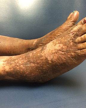
NEA tends to affect the lower limbs. However, there may be lesional involvement of the knees, thighs, genitalia, abdomen, buttocks, hands, and elbows, as well as the nails.3 Palms, soles, nails, and mucous membranes are generally spared. Patients typically report pruritus and may also complain of burning.
The manifestations have been divided into initial, well-developed, and late stages.4 Initially, scaly erythematous papules or plaques appear with a dark red-violet or eroded central region. Subsequently the lesions coalesce and become scaly with lichenification. Hyperpigmentation increases, and pustules may appear. In the late stage, hyperpigmentation remains, there may be superficial epidermal necrosis, and the lesions become thinner with possible crusting and erosions.
The histopathological features of NAE also vary by stage (Table).4 NAE lesions may undergo spontaneous remission and relapse.5
Etiology and pathophysiology
NEA is highly linked to HCV infection and may develop preceding onset of viremia. The population rate of HCV is relatively high in Egypt. Interestingly, no cases have been reported in Japan, where there is also a high prevalence of HCV, and NAE has been reported in the absence of HCV.3 It has also developed following vaccination for hepatitis B infection and after treatment with rivaroxaban.6,7
The pathophysiology of NAE is yet to be fully elucidated. It may involve hepatic dysfunction leading to metabolic abnormalities, particularly zinc deficiency, but also including hypoalbuminemia, hypoaminoacidemia, and hyperglucagonemia. Zinc deficiency has been reported in lesional and perilesional skin in patients with a normal serum zinc level.8
Differential diagnosis and work-up
Conditions to consider in the differential diagnosis of NAE include chronic eczema, psoriasis, necrolytic migratory erythema, lichen simplex chronicus, acrodermatitis enteropathica, and pellagra, although these alternative diagnoses can often be ruled out based on the absence of their hallmark clinical and laboratory features.
Necrolytic migratory erythema, a condition related to glucagonoma, tends to develop in intertriginous areas. In addition, affected patients will have elevated blood glucose and glucagon levels and likely will present with unintentional weight loss. Cutaneous lesions associated with pellagra usually develop on sun-exposed skin, and the patient’s history may include gastrointestinal symptoms. Laboratory testing should include serology for HCV infection, liver function tests, and zinc. Other tests to consider include serum glucagon, biotin, free fatty acids, and vitamin B3.
Management
Patients with NAE who test positive for HCV infection should receive antiviral treatment with interferon alpha-2b and/or ribavirin and should be referred to a gastroenterologist, hepatologist, or infectious disease specialist. Oral zinc supplementation (200 mg BID) is also indicated regardless of the patient’s zinc level. Zinc may provide therapeutic benefit as a treatment for NAE through multimodal mechanisms that relate to its reported anti-inflammatory, immunomodulatory, antiviral, and antioxidant properties.3 Consistent with published reports, the only patient I have seen with NAE achieved complete resolution when treated with oral zinc supplementation.
Other treatments for NAE reported in the literature include topical tacrolimus, vitamin B1 and vitamin B6, and phototherapy.3,9 Topical, intralesional, and systemic corticosteroid treatment has also been described, but without consistent benefit.2,3 Tinea pedis should be identified and treated in any patient with a dermatitis involving the feet.
Patients with this rare condition should be counseled that while the condition may clear, there can be residual scarring and pigmentary changes, and the lesions may recur.²
References
1. el Darouti M, Abu el Ela M. Necrolytic acral erythema: a cutaneous marker of viral hepatitis C. Int J Dermatol. 1996;35(4):252-256.
4. Abdallah MA, Ghozzi MY, Monib HA, et al. Histological study of necrolytic acral erythema. J Ark Med Soc. 2004;100(10):354-355.
3. Inamadar AC, Shivanna R, Ankad BS. Necrolytic acral erythema: current insights. Clin Cosmet Investig Dermatol. 2020;13:275-281.
2. Nutan F. Necrolytic acral erythema. June 25, 2020. https://emedicine.medscape.com/article/1098243-overview#a4. Accessed October 14, 2020.
5. Bentley D, Andea A, Holzer A, Elewski B. Lack of classic histology should not prevent diagnosis of necrolytic acral erythema. J Am Acad Dermatol. 2009;60(3):504–507.
6. Pernet C, Guillot B, Araka O, Dereure O, Bessis D. Necrolytic acral erythema following hepatitis B vaccination. Br J Dermatol. 2014;171(5):1255–1256.
7. Pathania YS, Budania A. Rivaroxaban induced necrolytic acral erythema. Postgrad Med J. 2019;1.
8. Najarian DJ, Lefkowitz I, Balfour E, Pappert AS, Rao BK. Zinc deficiency associated with necrolytic acral erythema. J Am Acad Dermatol. 2006;55(5 Suppl):S108–S110.
9. Manzur A, Siddiqui AH. Necrolytic acral erythema: successful treatment with topical tacrolimus ointment. Int J Dermatol. 2008;47(10):1073-1075.
