
From the files of Seemal Desai, MD, with Cheryl Guttman Krader
CASE HISTORY
An Indian woman presented with a 1-year history of hair loss and brown spots on her scalp. She had recently moved from India to Dallas, Texas and said she had seen a dermatologist for her problem once before.
On history, the patient was asymptomatic. She denied using any hair dyes or chemicals, starting any new medications, or having any illnesses or infectious exposures preceding the onset of her problem.
Examination showed widespread areas of hair loss with diffuse thinning. Dense, deep hyperpigmentation with a purplish hue was noted at the frontal hairline. Body examination revealed dense, velvety reticulated pigmentation and discoloration in the axilla and patchy pigmentation in a guttate pattern along the lateral ribcage and abdomen. A complete examination was not possible, however, because the patient declined to fully undress.
Laboratory tests were ordered and punch biopsies taken from the right lateral scalp and mid-abdomen. An antinuclear antibody test was negative, and results from both the complete blood count and complete metabolic panel were unremarkable. The biopsy showed dense dermal melanophages and possible resolved interface dermatitis. Mild vacuolar change was noted in one punch. There was no follicular plugging or active inflammation.
What is your diagnosis?
DISCUSSION
The patient was diagnosed with lichen planus pigmentosus (LPP) based on the pathology report. Although LPP is considered to be rare, dermatologists can anticipate seeing this condition more often because cases among people with skin of color, particularly of Indian and Middle Eastern descent, appear to be increasing at an almost epidemic rate. Whereas 10 years ago, only a rare patient presented with LPP, currently such cases are seen daily in practices serving a large population of patients of southeast Asian origin. Although there is no known exact trigger for LPP, anecdotally, pigmentary disease experts suggest one possible explanation for this phenomenon is that it is related to an epitope-like inflammation in the immune system induced by exposure to the presence of lead as a contaminant in certain spices that are used for cooking in southeastern Asian, Indian, and Pakistani cuisine.
The purple color of the pigmentation seen in this patient was a clue to the diagnosis. Otherwise, her clinical features, which included dense pigmentary changes involving the scalp and nonscarring alopecia, were atypical for LPP.
The absence of complete areas of hair loss ruled out alopecia areata. The combination of hair loss, hyperpigmentation, and atrophy suggested chronic cutaneous lupus erythematosus, which was excluded based on the laboratory and biopsy findings. Contact dermatitis of the scalp caused by paraphenylenediamine found in henna-based hair dyes was also suspected, as well as the possibility that hair dye use was the cause for the scalp pigmentation while the hair loss was coincidental. However, the patient denied use of any hair dye. Fixed drug eruption was also ruled out based on her history.
Lichen planus pigmentosus is a member of a family of acquired macular hyperpigmentary disorders of uncertain etiology that also includes ashy dermatosis (AD), erythema dyschromicum perstans (EDP), and idiopathic eruptive macular pigmentation (IEMP). All these disorders present with ashy pigmentation caused by melanophages in the dermis (Figure).
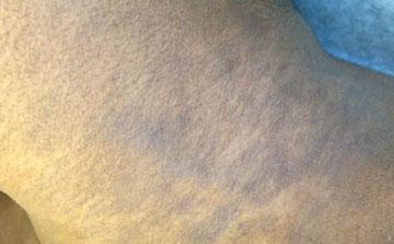
Figure. Lichen planus pigmentosus on left lateral neck showing classic hyperpigmented violaceous-to-dusky-gray patches in a reticulated configuration. Image courtesy of Seemal P. Desai, MD. [DR DESAI SAID : PLEASE watermark as copyright Seemal R. Desai MD or whatever format so that the readers know it cannot be reproduced.]
LPP, AD, and EDP can share features on histology and have been considered by some authors to represent a spectrum of a single entity. As highlighted in a report from an expert forum convened to develop a consensus on terminology for these acquired hyperpigmentary disorders, however, LPP and AD/EDP are distinct diseases that are differentiated clinically based on morphology and distribution (Table). Correct diagnosis is important for therapeutic decisions.
Table. Clinical and histological features of LPP and AD/EDP1
| Feature | LPP | AD/EDP |
| Morphology | Small (1-2 cm) macules | Large (>5 cm) macules surrounded early after development by an erythematous border |
| Predominant distribution | Head and neck in most cases with downward spread; flexural areas | Trunk |
| Histology | Dermal melanophages, possible mild interface dermatitis | Burned-out pigmentation, post-inflammatory pigmentary alteration, dermal melanophages, possible interface change |
The names AD and EDP may be used interchangeably, although EDP is preferred because of the negative social connotations associated with use of “ashy dermatosis” to describe a condition in patients with skin of color. Ashy dermatosis/EDP develops in patients of all ages and presents with large (>4-5 cm) nummular coalescing patches of ashy blue-gray lesions involving the trunk. The lesions appear first on the upper chest and back and then spread downward. The name EDP derives from the presence of an erythematous border that surrounds the macules during the early active phase of the disorder. Based on their color and erythematous border, lesions of EDP resemble those of a fixed drug eruption, although the latter condition usually is associated with induration and swelling.
In LPP, the pigmented macules are smaller (1–2 cm). They classically appear on the forehead, temples, ears, neck, and flexural areas, particularly the axillae, and can involve the trunk as they spread downward. The nomenclature for this entity is slightly confusing. Based on histological findings and reports of a clinical association with LP in some patients, LPP has been described as a variant of LP.1 However, only a minority of patients with LPP have a history of or existing LP . LP should be considered in the differential diagnosis for patients with acquired macular hyperpigmentation, but patients who have pruritic papules with residual hyperpigmentation should be diagnosed as having LP with post-inflammatory hyperpigmentation, and not LPP.
Idiopathic eruptive macular pigmentation is a self-resolving disorder that most often occurs in adolescents and young adults. The lesions are asymptomatic, small (0.5–2.5 cm) brown macules that typically appear on the neck, proximal extremities, and trunk and resemble residual lesions from guttate psoriasis.
Many modalities have been used to treat LPP. In my experience, low-dose isotretinoin (20–30 mg/day) continued for 6 to 8 months is highly effective. It was also identified as a promising treatment in a prospective open-label study including 32 patients.2 If the condition is diagnosed early and the biopsy shows evidence of inflammation, an initial tapering course of oral prednisone starting at 60 mg/day with bridging into isotretinoin is prescribed instead. Patients are also instructed about sun avoidance along with use of sunscreen, antioxidants, and topical lightening agents.
References
1. Kumarasinghe SPW, Pandya A, Chandran V, et al. A global consensus statement on ashy dermatosis, erythema dyschromicum perstans, lichen planus pigmentosus, idiopathic eruptive macular pigmentation, and Riehl’s melanosis. Int J Dermatol. 2019;58(3):263-272.
2. Muthu SK, Narang T, Saikia UN, et al. Low-dose oral isotretinoin therapy in lichen planus pigmentosus: an open-label non-randomized prospective pilot study. Int J Dermatol. 2016;55(9):1048-1054.
Disclosures
Dr. Desai reports no relevant financial interests.
