A 20-Year Intermittent, Mildly Pruritic Eruption
A healthy 42-year-old female patient presents with a two-decade history of an intermittent, mildly pruritic eruption involving the anterior chest. Self-treatment has failed. Patient history and physical exam are non-contributary. What’s your diagnosis?
By Ted Rosen, MD
Professor of Dermatology, Baylor College of Medicine, Houston, Texas
CASE HISTORY
An otherwise healthy 42-year-old ICU nurse presents with a two-decade history of an intermittent, mildly pruritic eruption involving the anterior chest. She has obtained, tried and failed self-treating with low potency topical steroids, several topical antifungals, triple antibiotic ointment, and topical crisaborole cream.
The patient is more concerned about the cosmetic aspect than about any medical ramifications, as the eruption has been present a long time.
Additional history, including past medical, family, and social history were entirely benign and non-contributory. Review of systems failed to disclose any significant complaints. The patient did not ingest any prescription medications with regularity, nor use any eye, ear, or nose drops. She denied all OTC medications, with the exception of daily multiple vitamins.
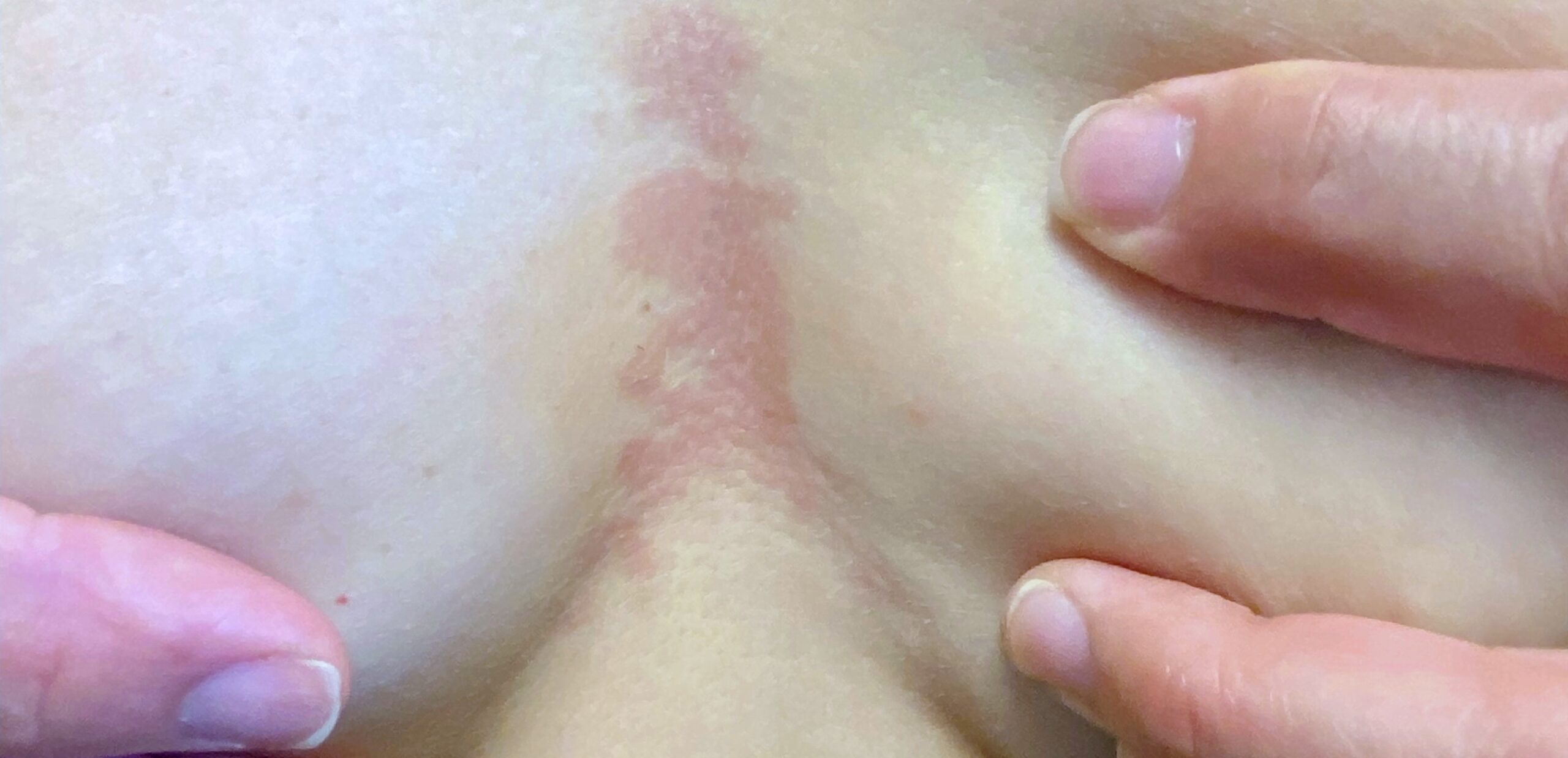
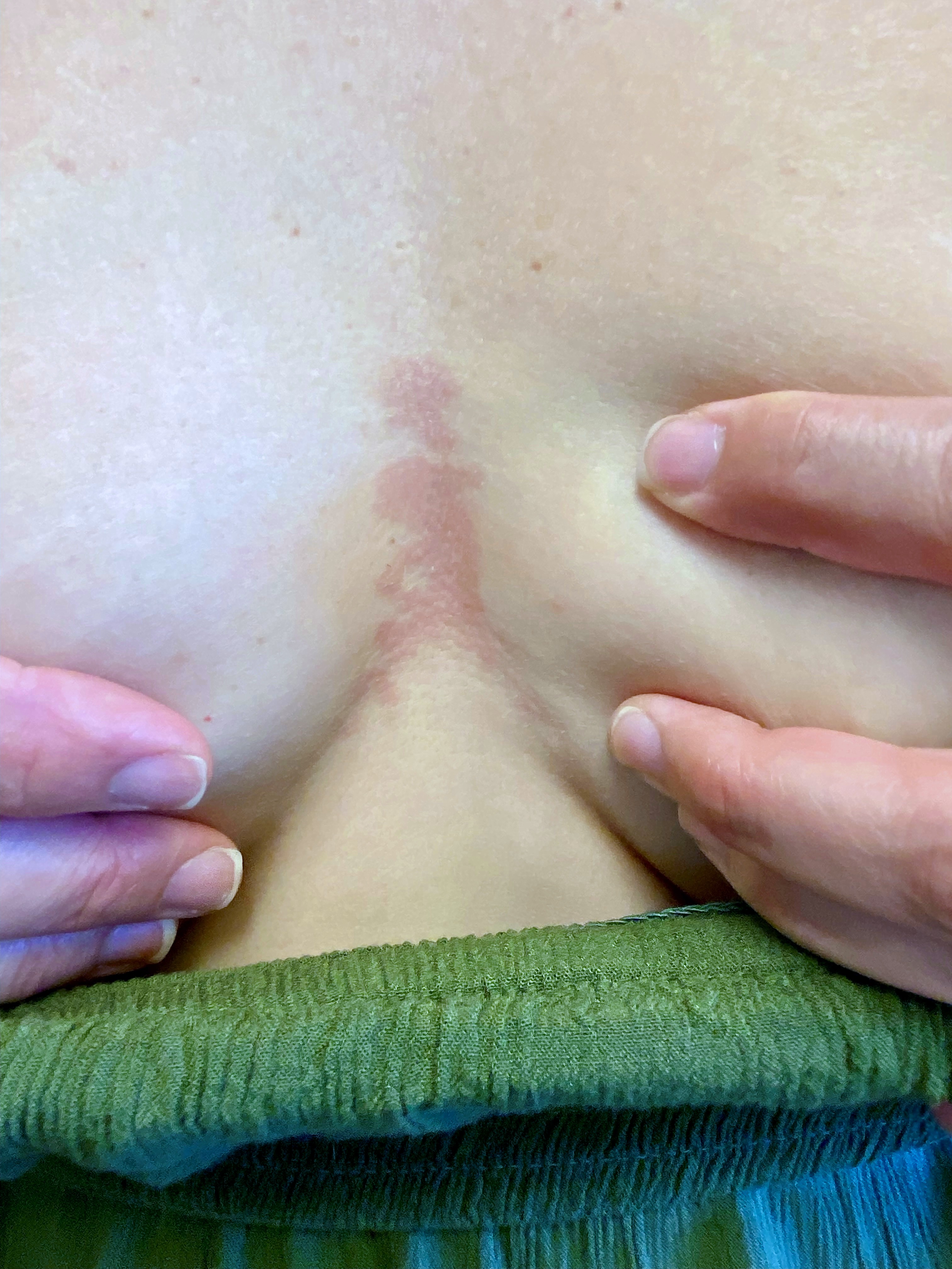
Physical examination revealed a well-defined area of slightly palpable, light-pink erythema located on the pre-sternal area, between the breasts. (Figure 1) The eruption was not tender to palpation. There was neither supraclavicular nor axillary adenopathy.
A recent chest radiograph was interpreted as normal. A brief full-body cutaneous examination failed to reveal any additional abnormalities or any areas similar to the one of concern. After obtaining informed consent, a punch biopsy was obtained, and the defect closed with 6-0 nylon sutures.
What is your diagnosis?
DISCUSSION
Histology revealed a normal orthokeratotic epidermis. Periodic acid–Schiff (PAS) stain did not reveal fungal elements in the stratum corneum. The dermis was notable for a superficial perivascular lymphocytic infiltrate with interstitial mucin visible on hematoxylin-eosin stained sections and verified by special stains. This pathology, along with the clinical morphology and history, is all consistent with reticular erythematous mucinosis (REM).
REM is a rare cutaneous disorder of uncertain etiology. There is a female predominance, with REM favoring women in the third and fourth decades of life. It has occasionally been linked to an antecedent viral infection, Hashimoto’s thyroiditis, and autoimmune diseases. REM has rarely also been associated with malignancy of the lung, breast, and colon. As was true in this case, the midline chest and back are the most typical locations.
REM resembles tumid lupus based upon histology, and some experts feel the two entities are identical. Our patient has neither signs nor symptoms compatible with lupus, and subsequent laboratory investigations (CBC, comprehensive metabolic panel, urinalysis, antinuclear antibody (ANA), anti-double stranded DNA, anti-Ro and La and anti-phospholipid antibody) were all normal or negative.
Treatment of choice remains administration of oral antimalarial medication; however, exposure to UVA-1 and 308 nm UVB (excimer laser) have proven successful in small numbers of cases. Asymptomatic disease of limited extent does not necessarily require intervention.
