Leaf-like Areas vs Streaks and Pseudopods
A patient in her fourth decade presents with a mole she first noticed 2 years ago. The mole has variegated color, border irregularity, and asymmetry. Initial impression is melanoma, but is it?
By Zaeem Nazir, MD, and Ashfaq A. Marghoob, MD
Dermatology Service, Department of Medicine, Memorial Sloan Kettering Cancer Center
New York, New York
CASE HISTORY
A patient in her 40s presents to the clinic for a new mole on her lower back, which she first noticed 2 years ago. She was planning to see a dermatologist but was unable to once the COVID pandemic started. She has no personal or family history of melanoma or non-melanoma skin cancer.
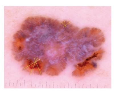
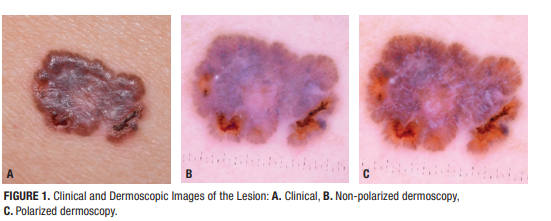
On clinical and dermoscopic exams, you observe the following:
Question: For this lesion, which dermoscopic feature is most diagnostic?
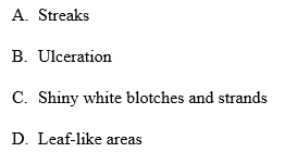
Answer: D. Leaf-like Areas
This patient has a lesion clinically suspicious for superficial spreading melanoma with variegated color, border irregularity, and asymmetry. While the initial impression has been a crucial driver for triage and diagnosis in medicine for centuries, anchoring bias can affect one’s subsequent judgement, even with evidence to the contrary. With a diagnosis of melanoma in mind, one may appreciate shiny white structures, radial streaks, blue-white veil, and regression structures on dermoscopy—all features associated with the diagnosis of melanoma.
What is your diagnosis?
DISCUSSION
A broader differential would also include pigmented basal cell carcinoma (BCC), as it can present similarly to melanoma. However, differentiating the two can be challenging as they are both associated with shiny white blotches and strands, brown dots, and atypical vascular structures. While the morphology of shiny white structures found in both have been hypothesized to help differentiate the two, present literature has confirmed that shiny white blotches and strands are associated with both BCC1 and nodular melanoma >2mm in thickness.3 In this particular case, the presence of a rolled border in this lesion provides strong evidence the lesion may be a pigmented BCC and not melanoma.
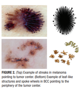
The key to this diagnosis is not to confuse the streaks associated with melanoma for the leaf-like structures associated with pigmented BCC. Streaks are defined as bulbous projections which must not be disconnected from the tumor or have an obtuse angle (>90) to the tumor edge4; they appear to radiate away from the tumor core and commonly point to the hyperpigmented center of a lesion (Figure 2). In contrast, leaf-like structures or spoke wheels are brown to gray-blue discrete bulbous extensions2 which converge focally, often towards the periphery of lesions that commonly have a hypopigmented center (Figure 2). The latter are highly diagnostic for BCC, with a specificity of 100%.
Appreciation for this key finding may help us identify other diagnostic structures for BCC, such as ulceration and shiny white blotches/strands which both have specificities of 95%.1,2 Other diagnostic structures to look for include blue-gray ovoid nests (specificity 99%), arborizing telangiectasia (92%), and spoke-wheel-like structures (100%).1
References
- Navarrete-Dechent C, Bajaj S, Marchetti MA, Rabinovitz H, et al. Association of shiny white blotches and strands with nonpigmented basal cell carcinoma: evaluation of an additional dermoscopic diagnostic criterion. JAMA Dermatol. 2016;152(5):546. doi:10.1001/jamadermatol.2015.5731.
- Menzies SW, Westerhoff K, Rabinovitz H, et al. Surface Microscopy of Pigmented Basal Cell Carcinoma. Arch Dermatol. 2000;136(8). doi:10.1001/archderm.136.8.1012.
- Sgouros D, Lallas A, Kittler H, et al. Dermatoscopic features of thin (≤2 mm Breslow thickness) vs. thick (>2 mm Breslow thickness) nodular melanoma and predictors of nodular melanoma versus nodular non‐melanoma tumours: a multicentric collaborative study by the International Dermoscopy Society. J Eur Acad Dermatol Venereol. 2020;34(11):2541-2547. doi:10.1111/jdv.16815.
- Menzies SW, Crotty KA, McCarthy WH. The Morphologic Criteria of the Pseudopod in Surface Microscopy. Arch Dermatol. 1995;131(4):436-440. doi:10.1001/archderm.1995.01690160064010.
- Tavoloni Braga JC, Scope A, Klaz I, Mecca P, et al. Melanoma mimicking seborrheic keratosis: An error of perception precluding correct dermoscopic diagnosis. J Am Acad Dermatol. 2008;58(5):875-880. doi:10.1016/j.jaad.2007.12.011.
Disclosures: No relevant disclosures.
