African American with Torso Lesions, History of Spontaneous Keloids
By Ted Rosen, MD, FAAD

CASE HISTORY
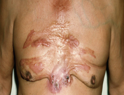
A 36-year-old African American male presented for evaluation of lesions involving the anterior torso, extending from the level of the clavicle to the uppermost abdomen, just inferior to the nipple line.
He had developed these spontaneous keloids at age 15 to 17. But other than being unsightly, the lesions had generally only been associated with mild, intermittent tenderness. Recently, a number of the keloids had become both pruritic and painful. Notably, the same symptomatic keloids had begun to exude purulent drainage. The longstanding asymptomatic lesions, however, were not demonstrating purulent discharge.
Past medical history was not contributory. Family history was positive (father and one brother) for spontaneous keloids of the chest. A recent biochemical panel and complete blood count were entirely within normal limits. Specifically, there was no leukocytosis.
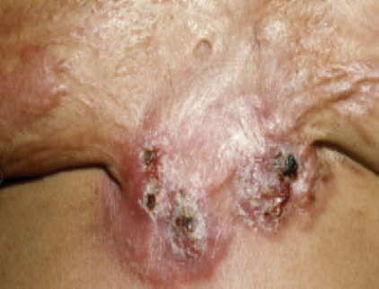
Physical examination revealed at least 13 distinct keloids located only on the anterior torso. Six areas of recent purulent exudation, now inflamed and covered by crusts, were identified
Pressure on any of these areas would elicit purulent discharge. There was no axillary adenopathy. Chest radiograph was normal. The remainder of the examination was unremarkable. Specifically, the patient was (and had been) afebrile.
Biopsy revealed a few foci of folliculitis within a classic dense collagen stroma, consistent with a keloid. Aerobic, anaerobic, mycobacterial and fungal cultures of expressed purulence from several lesions yielded no growth.
What is your diagnosis and how would you help him?
DISCUSSION
The diagnosis is suppurative keloidosis, a rare variant of spontaneous keloid formation. This entity consists of largely sterile purulence developing in previously asymptomatic keloids.
It has been estimated that about one-quarter of keloid patients may develop this phenomenon.1,2 Patients at increased risk for suppuration to develop are typically of African ancestry, male gender, with many keloids, and with a positive family history for keloid formation.
Clinical folliculitis may precede the presence of keloid suppuration, but cultures fail to yield anything other than occasionally normal skin flora. Since this is not an infectious process, it is suggested that use of antibiotic therapy is not appropriate. It is recommended that intralesional corticosteroid injections form the basis for therapy.1-3
This particular patient’s lesions responded nicely to intralesional injections of triamcinolone acetonide, 5-10mg/ml concentration. Because the aseptic process involved in suppurative keloidosis is likely similar to that involved in hidradenitis, it has also been suggested that use of TNF-alfa blockers might prove beneficial. In fact, one such instance has already been documented.4
References:
- Delaleu J, et al. Suppurative keloids: a complication of severe keloid disease. Int J Dermatol. 2021;60(11):1392-1396. doi:10.1111/ijd.15641.
- Olaitan PB. Keloids: assessment of effects and psychosocial-impacts on subjects in a black African population. Indian J Dermatol Venereol Leprol. 2009;75(4):368-372. doi:10.4103/0378-6323.53132.
- Novick NL, et al. Suppurative keloidosis in a black woman. J Am Acad Dermatol. 1986;15(5 Pt 2):1090-1092. doi:10.1016/s0190-9622(86)70270-3.
- Jfri A, et al. Association of hidradenitis suppurativa and keloid formation: A therapeutic challenge. JAAD Case Rep. 2019;5(8):675-678. Published 2019 Aug 2. doi:10.1016/j.jdcr.2019.06.001.
