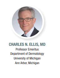
Dr. Charles N. Ellis explains why measuring epicardial adipose tissue volume when patients with psoriasis are undergoing computed tomography cardiac imaging might be important for assessing cardiovascular risk in these patients.

“Including measurement of epicardial adipose tissue volume (EAT-V) when patients with psoriasis are undergoing computed tomography (CT) cardiac imaging should be considered because it may provide valuable additional information for assessing cardiovascular risk,” said Charles N. Ellis, MD, co-lead author of a published study investigating EAT-V in psoriasis patients.1
“We know that patients with psoriasis have an overall increase in systemic inflammatory load that puts them at increased risk for future cardiovascular events, and therefore it is advisable to screen patients with psoriasis for cardiovascular risk factors. Although cardiac imaging is done based on clinical need and is not considered a screening test, we believe that EAT-V should be reported whenever a patient with psoriasis is undergoing a CT cardiac imaging test.”
“Measurement of EAT-V is a relatively simple, safe, reproducible test that might increase our ability to identify patients who are at high risk for coronary events so that we could focus prevention efforts on patients who need it the most. Risk awareness is the first step toward prevention,” he said.
What Is EAT and Why Does It Matter?
Epicardial adipose tissue lies between the myocardium and pericardium. It is metabolically active tissue that secretes proinflammatory and proatherogenic cytokines. The proximity of release of these deleterious compounds to the coronary arteries is believed to explain a documented association between EAT-V and coronary artery disease.
Although available research shows that psoriasis is associated with coronary artery disease as measured by the severity of coronary artery calcification, previous studies investigating the risk of future cardiovascular events and related mortality in patients with psoriasis have yielded variable results.
“Thus there exists an unmet need for simple clinical evaluations that help predict the risk of serious cardiovascular events for patients with psoriasis,” said Dr. Ellis.
EAT-V In Psoriasis Patients
Previous studies investigated EAT-V in patients with psoriasis and reported it was greater than in controls. However, most of these studies used single-location ultrasound to evaluate EAT thickness, which is only a surrogate measure of EAT-V. The studies also varied in their criteria for selecting patients with psoriasis and controls.
To further investigate the question of whether EAT-V is increased in patients with psoriasis, Dr. Ellis and colleagues conducted a cross-sectional, controlled study choosing subjects with psoriasis and controls from a convenience sample of patients seen at the University of Michigan Dermatology clinics. They focused on patients with severe chronic plaque psoriasis, defined as having >10% body surface area involvement and at least one episode of inpatient therapy or systemic treatment for psoriasis. Controls had no personal or family history in first-degree relatives of psoriasis or other rheumatologic diseases.
Clinic patients were excluded from the study if they were age <18 or >55 years, had any history of heart disease, were receiving TNF-alpha inhibitor treatment, had diabetes mellitus, were pregnant, or weighed over 320 pounds.
EAT-V was determined using 3-dimensional CT, which is considered to provide accurate and reliable quantification of EAT-V. Coronary artery calcification and coronary artery plaque and stenosis were also scored. All measurements were done by an experienced cardiothoracic radiologist who had no knowledge as to whether the subject had psoriasis.
The study included 25 psoriasis patients and 16 controls; 40 participants were White.
Comparisons of demographic and cardiovascular risk factors between the patients with psoriasis and controls showed that the only statistically significant difference was for EAT-V which was greater in the psoriasis group than in controls (91 vs 70 cm3; P =.04). There were no significant differences between the psoriasis patients and controls in either coronary artery calcification or plaque formation, likely due to selection criteria of relatively young age and absence of known heart disease.
Subgroup analyses of the data with patients divided by sex showed that EAT-V was statistically higher in the psoriasis patients compared to controls only among males.
Addressing the Limitations
As a small cross-sectional study performed in a convenience sample that included only patients with severe psoriasis, the research has several limitations, said Dr. Ellis.
“Because of our study’s small sample size and lack of racial diversity, future areas of research include seeing if our findings are confirmed in a larger patient population and if EAT-V differs between patients with psoriasis and controls among non-Caucasians,” he said.
“In addition, we believe further investigation is needed to explore whether female psoriasis patients have increased EAT-V. Although we found such a difference existed in a numerical sense, we could not prove it statistically. The explanation may lie partly in the fact that women are generally smaller and have less EAT-V than men to begin with. However, if more women are studied, I expect the results will show that EAT-V is increased in women with psoriasis just as we found in men.”
“Whether elevated EAT-V is a result of increased general inflammation or is an independent cause of the increased cardiovascular risk, or both, is another opportunity for further study. We hope our colleagues in dermatology and radiology will conduct the research needed to expand on our initial findings,” said Dr. Ellis.
Dr. Ellis predicts that demand for quantifying EAT-V in patients with psoriasis will increase as the significance of this metric becomes more widely recognized.
“Moreover, the rise in demand for EAT-V measurement will likely result in EAT-V measurement becoming more automated because it might motivate manufacturers of CT platforms to develop software capable of reporting the information without human input from the radiologist. However, EAT-V will not become a standalone test for prognosticating cardiovascular risk because obtaining the measurement requires patients to have clinical indications for the CT cardiac imaging that allows calculation of EAT-V,” he said.
Reference
Ellis CN, Neville SJ, Sayyouh M, et al. Epicardial adipose tissue volume is greater in men with severe psoriasis, implying an increased cardiovascular disease risk: A cross-sectional study. J Am Acad Dermatol. 2022;86(3):535-543. doi:10.1016/j.jaad.2021.09.069.


