Large, Painless, Thin-Walled Blister Ruptures, Then Many Smaller Bullae Appear
By Ted Rosen, MD, FAAD
CASE HISTORY

A 78-year-old male being treated for rheumatoid arthritis with adalimumab and low dose prednisone (10 mg daily) developed a solitary painless red plaque on the abdomen, which, in 12 hours, evolved into an eschar.
At that point, the patient felt unwell and became febrile and reported to the emergency department. The lesion was recognized as ecthyma gangrenosum, typically (about 75%) associated with pseudomonas septicemia. He was admitted and started on appropriate intravenous antibiotic therapy (piperacillin/tazobactam and levofloxacin). This intravenous regimen was continued for two weeks.
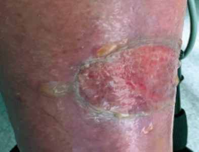
On day 12, however, the patient developed a large, painless, thin-walled blister on his left shin. This initial blister ruptured but was followed by the appearance of multiple smaller tense bullae close to the site of the first lesion.
Dermatology was then consulted for the first time and noted that both legs were grossly swollen and erythematous. Prior chart notes had commented on pre-existing venous insufficiency and associated stasis dermatitis. A biopsy was done on one of the small bullae and revealed a subepidermal split with a paucicellular lymphocytic perivascular infiltrate and widened spaces between collagen bundles. All tissue stains for microorganisms were negative.
What is your diagnosis?
DISCUSSION
The patient has fairly classic edema bullae, also known as edema blisters.
This non-infectious, non-immunologic cause of blister formation occurs mostly in elderly, immobile individuals during periods of increased interstitial pressure.
Edema bullae are medium to large sized, with a thin roof which frequently breaks.
The etiopathogenesis of this phenomenon is when capillary filtration develops too rapidly for compensatory lymphatic drainage.
Underlying causes for this physical phenomenon include hepatic, renal, or cardiac failure or simple fluid overload (often iatrogenic), particularly in the setting of venous insufficiency. This particular patient had a history of long-standing venous insufficiency and stasis dermatitis, and then was subject to intravenous fluids for two weeks. This appropriate but unfortunate fluid overload led to such intense peripheral edema that bullae formation took place.
Treatment of edema bullae is directed toward reducing the fluid overload and tissue edema. Stopping the intravenous fluid as soon as possible was a critical step. Elevation of the affected extremity was another maneuver utilized to facilitate the removal of edema fluid from the blister-bearing site.
Administration of diuretics may also be considered. Lymphatic obstruction may be sought when there is no prior history of venous disease, and any organ failure which is associated with edema should be corrected as feasible.
The key point is to recognize this relatively benign condition for what it is, initiate steps to reduce edema fluid, and avoid treatments for immunobullous disorders. A biopsy may certainly help distinguish between edema bullae and blisters due to pemphigus and pemphigoid, although biopsy of an edematous extremity may lead to a slowly healing wound.