PARPi-FL, a topical fluorescent molecular contrast agent, may detect basal cell carcinoma through intact skin in as little as five minutes, according to new preclinical research in ex vivo human tissues.
The findings are published in the August 2025 issue of The Journal of Nuclear Medicine.
The new data confirmed that PARPi-FL is non-toxic to the skin and does not cause systemic side effects, making it a potential one-stop-shop for diagnosis and management of basal cell carcinoma, the researchers report.
Emerging nonsurgical therapies for early basal cell carcinomas could be delivered at the bedside, but they require noninvasive diagnostic tools with high accuracy, says study author Manu Jain, MD, a Research Pathologist and Optical Imaging Specialist in Fermatology at Memorial Sloan Kettering Cancer Center in New York, NY, in a news release.
Research Goals
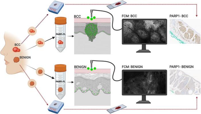
The study evaluated the optimal dose, application time, and diagnostic performance of PARPi-FL using ex vivo human tissues, including specimens from plastic surgery, Mohs surgery (basal cell carcinomas), and fresh excisions (benign and basal cell carcinomas). Researchers also investigated the feasibility of topical application using gauze and real-time in vivo imaging with a commercial reflectance confocal microscopy (RCM) device in tumor-bearing mice. Preclinical toxicology studies were conducted to determine safety as well.
A minimal topical dose of 10 M applied for two to five minutes via gauze achieved sufficient dermal penetration. PARPi-FL generated a strong fluorescent signal in basal cell carcinoma lesions with significantly weaker signals in benign tissues, supporting its diagnostic capability. Furthermore, preclinical data confirmed that PARPi-FL is safe for topical application, adds Ashish Dhir, PhD, DABT, Senior Pharmacology and Toxicology Manager in the Office of Entrepreneurship and Commercialization at Memorial Sloan Kettering Cancer Center in New York.
Incorporating this targeted dye into in vivo imaging could significantly improve diagnostic accuracy, reduce unnecessary biopsies of benign lesions, and enable timely, noninvasive treatment for basal cell carcinoma, notes Dr. Jain. Importantly, since PARP1 is also overexpressed in melanoma, these results support the feasibility of extending this approach to melanoma, offering a promising tool for distinguishing malignant from benign pigmented lesions without biopsy.
PHOTO CAPTION: PARPi-FL fluorescent confocal microscopy imaging for basal cell carcinoma detection.
PHOTO CREDIT: Image created by Manu Jain and Ashish Dhir, Memorial Sloan Kettering Center, NYC, USA.
