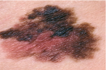
The activity of macrophages may be used to predict whether a melanoma patient will respond to immunotherapy, according to a study in JCO Oncology Advances.
For the study, researchers examined novel biomarkers that may identify melanoma patients more likely to respond to talimogene laherparepvec (TVEC).
TVEC is a modified oncolytic virus that is injected into melanoma directly to stimulate an immune response. It previously has been used in advanced melanoma; however, this study was the first to examine its potential to treat high-risk stage II melanoma patients upfront.
It was conventionally thought that TVEC worked by activating T cells. However, the team found that pre-existing and post-treatment T cell populations did not have association with treatment responses. Instead, they found that changes in the macrophages correlated to which patients responded to the treatment and which did not. Additionally, previous research has monitored amounts of protein indicators such as programmed death-ligand 1 (PD-L1) and at the genes involved in T cells to assess whether immunotherapy is effective.
However, this latest study shows these techniques do not accurately predict which patients will respond to treatment.
The researchers used a method called iFRET, which monitors protein activation instead of simply measuring the amounts of protein present. They found that T cell presence showed no consistent trends to viral stimulation or tumour response before and after the treatment, but there was a heavy infiltration of macrophages after treatment in responding patients, associated with very high activation across immune checkpoint regulators.
“We know that people respond to immunotherapy very differently – in some cases the tumours shrink, and in others, sadly the patients do not survive,” study author Professor Banafshé Larijani, Department of Life Sciences and Director of the Centre for Therapeutic Innovation at the University of Bath in Bath, U.K., in a news release. “Our findings show that it’s not enough to simply look at T cell activity, instead it’s imperative to look at the whole immune response environment in detail to predict how a patient will respond to different treatments. Our results suggest that, in non-responding patients, we should be targeting these macrophages to reprogramme the tumour immune environment.”
She continues, “We hope our research will enable clinicians to make important decisions over which patients would be better served by surgery or immune checkpoint blockade by immunotherapy.”
Amanda Kirane, MD, PhD, Director of Cutaneous Surgical Oncology Department at Stanford University School of Medicine in Stanford, CA, who led the clinical part of the study, adds: “This study is highly informative in establishing a connection between pre-existing innate immune functions and ability to respond to immune-stimulating drugs.
“It also strongly supports emerging evidence that there may be biological differences in patients more likely to respond to this kind of immunotherapy – oncolytic viruses – versus other types that target immune checkpoint regulators.
“Lastly, it extends new and important context to the disconnect between measuring PD-L1 protein values as a clinical biomarker and protein activity in the tumour.
“The added information of iFRET-based immune activity measurements may offer the critical missing link of why current biomarkers have failed to yield a usable test to aid patients in treatment decision-making.”
Next, the team aims to characterize all cells contributing to the immune checkpoint interaction, which will further improve patient stratification and therefore tailoring of personalized medicine.


