Researchers have recreated the structure of wrinkles in biological tissue in vitro, uncovering the mechanisms behind their formation.
Their findings were published in Nature Communications.
Professor Dong Sung Kim’s team from the Department of Mechanical Engineering at POSTECH developed an epithelial tissue model composed solely of human epithelial cells and extracellular matrix (ECM). By combining this model with a device capable of applying precise compressive forces, they successfully recreated and observed wrinkle structures in vitro that are typically seen in the gut, skin, and other tissues in vivo.
This allowed them, for the first time, to replicate both the hierarchical deformation of a single deep wrinkle caused by a strong compressive force and the formation of numerous small wrinkles under lighter compression. The team also discovered that factors such as the porous structure of the underlying ECM, dehydration, and the compressive force applied to the epithelial layer are crucial to the wrinkle formation process.
Their experiments revealed that compressive forces deforming the epithelial cell layer caused mechanical instability within the ECM layer, resulting in the formation of wrinkles. Additionally, they found that dehydration of the ECM layer was a key factor in the wrinkle formation process.
These observations closely mirrored the effects seen in aging skin where dehydration of the underlying tissue layer leads to wrinkle development, providing a mechanobiological model for understanding wrinkle formation.
“We have developed a platform that can replicate various wrinkle structures in living tissue without the need for animal testing,” says Professor Kim in a news release. “This platform enables real-time imaging and detailed observation of cellular and tissue-level wrinkle formation, processes that are difficult to capture in traditional animal models. It has wide-ranging applications in fields such as embryology, biomedical engineering, cosmetics, and more.”
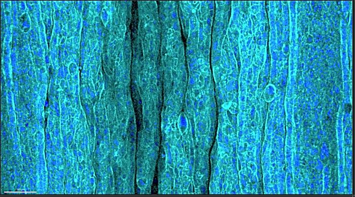
PHOTO CAPTION: Image of wrinkled epithelium on the ECM hydrogel layer in response to compression
PHOTO CREDIT: POSTECH
